 High-speed low-light in vivo two-photon voltage imaging of large neuronal populations
– nature methods (published: )
For years, scientists have been developing optical imaging as the non-invasive approach for recording neuronal electrical signals necessary for understanding brain function. Our researchers at The John B Pierce Laboratory and the Yale School of Medicine (Drs. Jelena Platisa and Vincent Pieribone) collaborated with colleagues at Boston University (Drs. Jerry Chen and Lei Tien Laboratories) to create a toolbox that pushes the limits on the duration of imaging studies in the living brain.
The work published today in the journal Nature describes the multifaceted system that couples a novel fluorescent protein-based probe (SpikeyGi), a super-fast multi-photon microscope (SMURF), and a data processing package (DeepVid) for the sustainable recording of spiking activity in hundreds of neurons in the living brain.
The study addresses several issues limiting the broader adoption of optical voltage imaging in the neuroscience community. First, the novel probe SpikeyGi detects neuronal electrical spikes with amplified sensitivity showing a spike-dependent increase in green fluorescence intensity. Unlike many other voltage indicators, SpikeyGi is compatible with multi-photon microscopy, the method of choice for recording neurons in the deeper regions of intrinsically opaque brain tissue. However, traditional multi-photon approaches have slow imaging rates incompatible with the sub-millisecond neuronal spike kinetics.
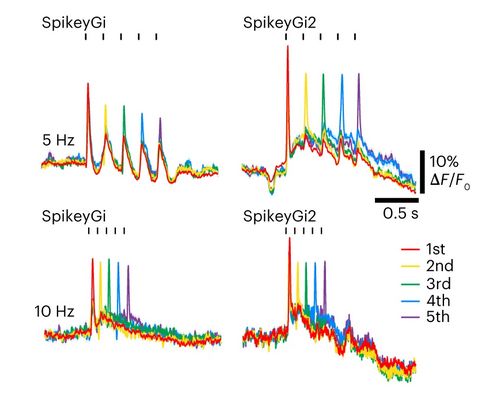
SpikeyGi and SpikeyGi2 performance during in vivo two-photon population imaging.
A novel super-fast two-photon microscope design, SMURF, uses spatial and temporal multiplexing of the laser beams to achieve unprecedented kilohertz imaging speeds from a large field of view. Finally, the self-supervised deep learning framework DeepVID enables the de-noising of the voltage imaging data acquired at low-light conditions. Combining these tools creates a unique framework for imaging brain electrical signals at spatial and temporal scales relevant to physiological studies.
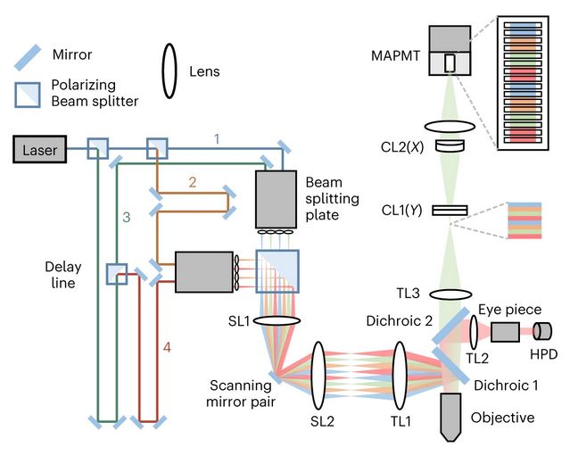
Design of SMURF two-photon microscope.
Click here for link to publication.
This study was funded by grants from the National Institutes of Health and DARPA.
The other authors of this study are Xin Ye, Allison Ahrens, Chang Liu, Ichun Anderson Chen, Ian Davison, Lei Tian, and Jerry L Chen.
